• Housekeeping!

    I just wanted to apologize for missing last week’s regularly scheduled post. I have been travelling and did not have time to work on my blog. So… yeah, sorry about that. I’ll try to plan ahead better next time.

    Before we start, I also wanted to let you know that I will be coming out with a new post every 2 weeks from now on, instead of every week. This is because I now have less free time than I did before (I got a job, lol). But don’t worry, we still have lots to talk about. Right then, to the post!

    More about chamomile flowers than you ever needed/wanted to know:

    I’ve been growing a chamomile plant in a little hydroponics unit sitting on a table indoors. It has been fun watching it grow, but recently I’ve become very interested in the flowers.

    Like sunflowers, chamomile is a member of the Asteraceae. Members of this family have inflorescences – that is, clusters of many flowers – which together resemble a single flower. There are two kinds of flowers within the inflorescence: Ray flowers and disc flowers. In chamomile, ray flowers have large white petals and disc flowers have comparatively tiny petals. You can see an example of a chamomile inflorescence in Figure 1 below.
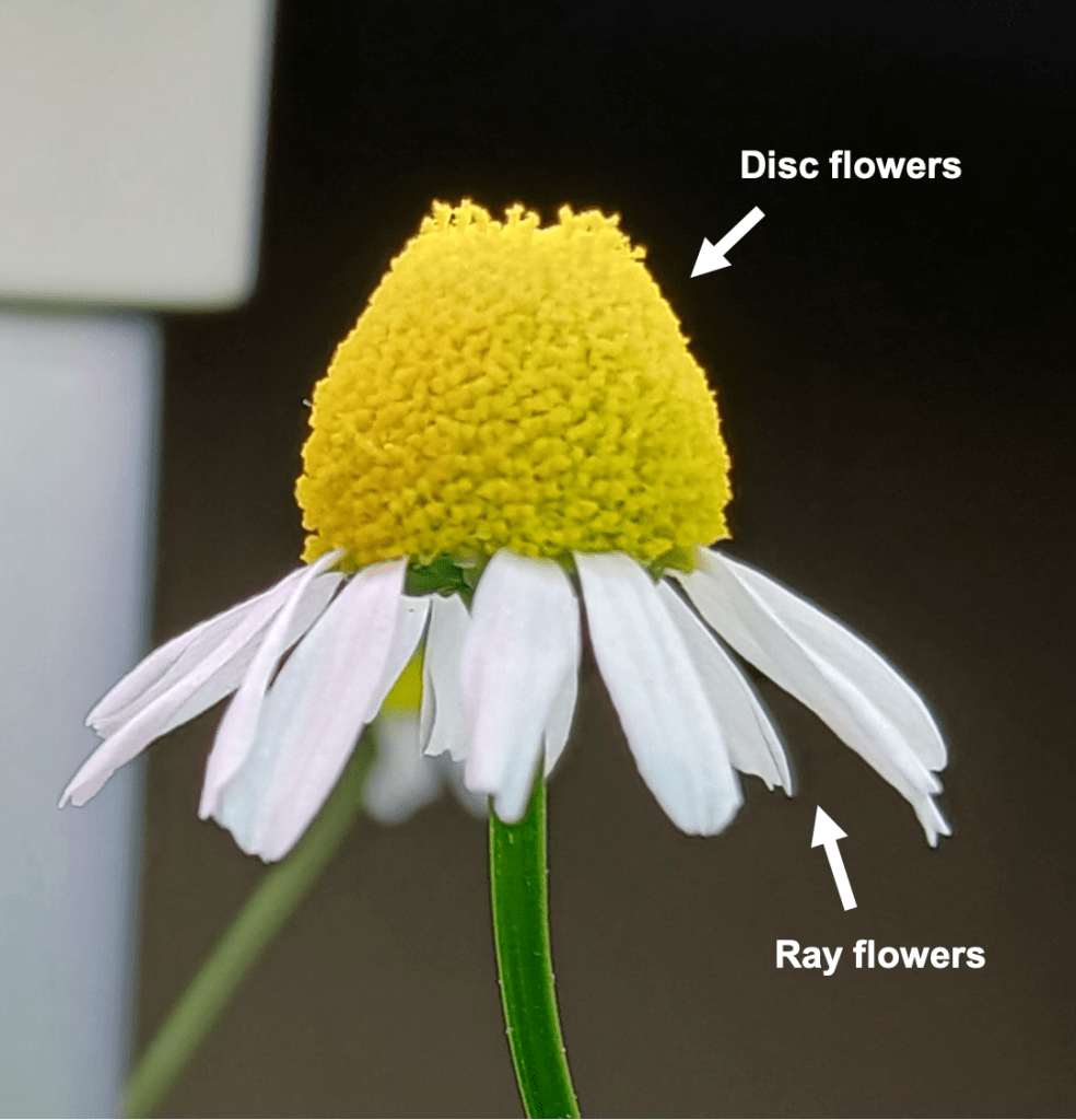
    Figure 1. Chamomile inflorescence, showing the location of ray flowers and disc flowers.

    You might be looking at this picture and thinking: “I don’t see any flower-shaped things in there. It just looks like one big flower.” That’s fair. So, let’s take a look at the ray and disc flowers under the microscope! Figure 2 shows a side view of a disc flower. The flower is so tiny that you can see the individual cells which make it up. We can see the petals, the stigma (a female reproductive part), and even scattered pollen grains.

    Figure 2. A chamomile disc flower at 100X magnification and blown up further to a larger size.  

    In Figure 3, we can see a ray flower. The structure of the female reproductive organs is similar to the disc flower; The stigma can be seen here, too. However, chamomile ray flowers are pistillate, meaning that they lack the male reproductive structures [6]. Neat! Furthermore, the petals are fused into a single large, flat plane that is comparatively enormous in size (I had to cut it off to fit the flower under the microscope).
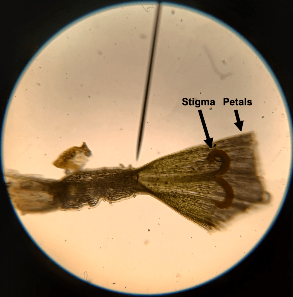
    Figure 3. A chamomile ray flower (with petals cut off) at 100X magnification. The fused petals are enormous and would have extended a long way to the right, had they not been severed.

    Check this out!

    This is all very fascinating, of course. But the REALLY cool thing about chamomile flowers is that they can move!

    No, really! I’m not crazy…. They can move – just very slowly. Now, I’ve been growing my chamomile plant in day-night light cycles. During the day, the petals of the ray flowers are erect and point out sideways. At night, the petals of the ray flowers hang downwards. To illustrate this, I photographed the same 2 flowers over a couple days to show this movement in action – see Figure 4 below.

    Figure 4. Daily petal movements of chamomile ray flowers. This image series follows the same pair of flowers across 2 days.

    While petal movements in chamomile are certainly striking, I should point out that daily rhythmic movements are actually quite common in plants. For example, if you’ve ever kept Oxalis triangularis (a common houseplant), you might also notice that the leaves and flowers move up and down, depending on the time of day (See Figure 5 below). Similarly, the leaves of the model plant Arabidopsis thaliana also move up and down depending on the time of day [2], though this movement is somewhat less striking than in Oxalis and involves a different organ (the petioles as opposed to pulvini within leaves themselves).

    Figure 5. Daily movements in an Oxalis kept as a houseplant. Left: The plant during the day. Right: The plant at night. These are my photos, but not my plant.

    In Oxalis and Arabidopsis, these rhythmic movements are controlled by the circadian clock [2][3]. The circadian clock is an intrinsic biological rhythm which controls daily biological cycles. You have a circadian clock which controls your sleep patterns (this is why you sleep at roughly the same time each day) and various aspects of your metabolism. Plants also have a circadian clock which controls rhythmic movements, among other things.

    Time to get a new watch…

    One defining characteristic of circadian rhythmics is that they persist when organisms are moved from cycling to constant conditions [3]. For example, when Arabidopsis is shifted from day/night light cycles to constant light conditions, up-and-down leaf movements continue on a roughly 24-hour cycle [2]. With this in mind, I asked myself whether the flower movements in chamomile were also controlled by the circadian clock.

    To test this, I severed some chamomile inflorescences, gave them some water, and put them into constant dark conditions (in my closet). I harvested two inflorescences at 4 PM (daytime), which roughly corresponds to the time when the ray flower petals are most erect. I also harvested two inflorescences at midnight. At this time, the ray flower petals hang loosely. (For your reference, my lights are kept on from 6 AM to 11 PM). Then, I tracked the movement of the flower petals over the next couple of days.

    Hypothesis: If the flower movements are controlled by circadian rhythms, then the petals should continue to move up and down in constant dark conditions.

    Great! We’re all set. So… what actually happened? The results actually did not support my hypothesis. As you can see in Figure 6, the inflorescences harvested during the day drooped after being moved to constant dark, and did not re-open. Meanwhile, the inflorescences harvested at midnight remained drooped throughout the entire experiment.
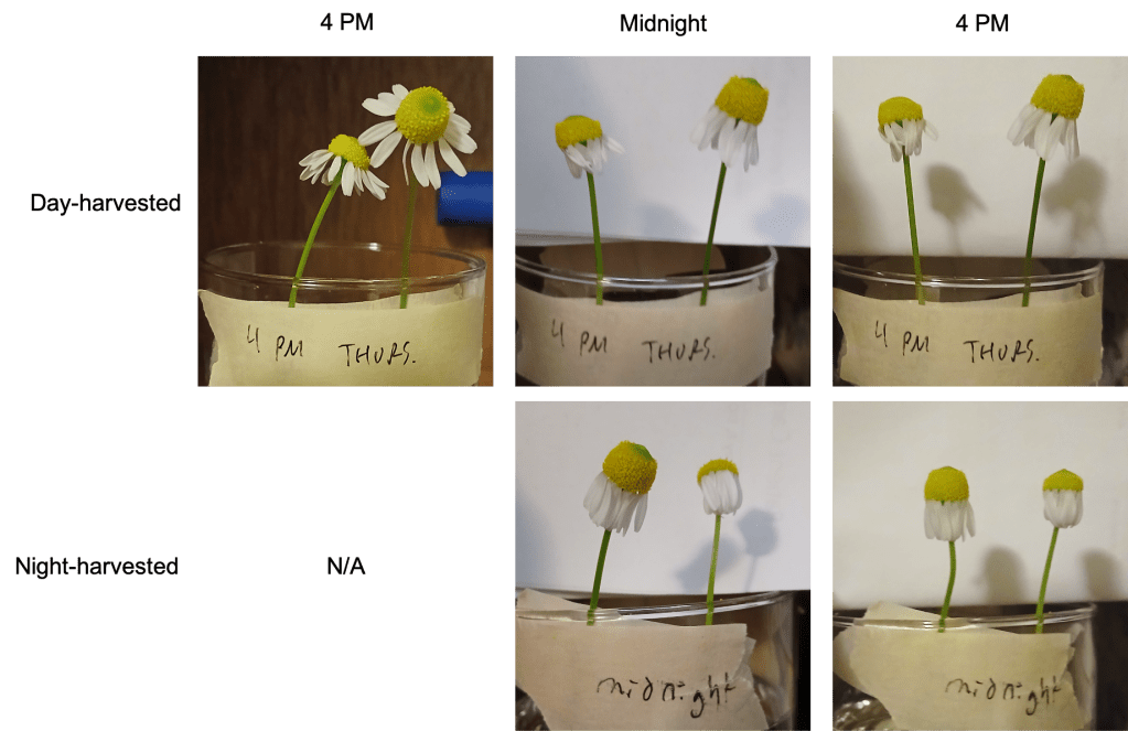
    Figure 6. Petal movements in chamomile flowers kept in constant darkness. The top row of images shows flowers harvested at 4 PM, during the day. The bottom row of images shows flowers harvested at midnight. Images in each row are in chronological order from left to right.

    Why did this happen? I don’t know, but I’ve thought of a couple possible explanations. Let’s discuss.

    POSSIBLE EXPLANATION 1: Flower movements in chamomile are not controlled by the circadian rhythm, but are rather directly controlled by present light levels.

    I think this is unlikely for a couple of reasons. First, I observed that the petals on my chamomile plant tend to start drooping at the end of the day, BEFORE the lights go off. In other words, they are anticipating an impending change in light conditions. This would not happen if petal movements were determined by light alone.

    I also tested this theory directly. To do this, I took my chamomile inflorescences that had been sitting in the dark for a few days (and were super droopy) and moved them back into the light. If light is necessary and sufficient to cause petals to stand up, then I would expect the petals in my cut inflorescences to do just that. They appeared to make what seemed to be a half-hearted attempt to stand up after many hours of light exposure, but ultimately did not move much. Interesting.

    POSSIBLE EXPLANATION 2: The act of severing inflorescences from the rest of the chamomile plant results in a loss of circadian petal movement. I can think of two possible reasons for this. One possibility is that petal movements are controlled remotely by a circadian oscillator in another part of the plant (the leaves, for instance). Another possibility is that petal movement requires a specific nutrient, which severed flowers in water would be unable to acquire.

    Sadly, I still haven’t had the time to test these ideas. This is because I cut off all of my chamomile flowers for my previous experiment (and I didn’t have many to begin with!). However, when my plant recovers and generates more flowers, I should be able to do a small experiment. If rhythmic petal movements require that the inflorescence be attached to the rest of the plant, then these movements should persist if the whole plant is transferred to constant dark conditions. I’ll let you know when the results of that test become available.

    Gene of the week:

    Tragically, I was not able to find any research about the genetics underpinning flower petal movements in chamomile or in a closely related species. Therefore, I have been forced to choose a Gene of the Week which is only tangentially related to what we were talking about. I still think you’re going to like it, though.

    This week’s Gene of the Week is ELF3. Funny name, eh? ELF3 stands for “EARLY FLOWERING 3”, because Arabidopsis plants lacking a functional copy of this gene flower early [4]. … I guess that makes sense. The ELF3 protein is an important component of the circadian clock in Arabidopsis. Without it, plant movements and other circadian-regulated processes are arrhythmic in constant light [5]. Interestingly however, elf3 mutant Arabidopsis plants still maintain rhythmicity in constant dark conditions [5].

    What does the ELF3 protein do? Famously, it is part of a larger protein complex (called the Evening Complex) which controls the expression of other genes [8]. As its name suggests, the Evening Complex is most active in the evening. Very generally, the Evening Complex helps to ensure that daily oscillations in gene expression occur at the correct time. Interestingly, recent research has also shown that ELF3 also has functions independently of the evening complex [7]… so it does rather a lot!

    Now, the BIG question: Does chamomile have an ELF3 gene? Well, my favorite ortholog database (OMA: https://omabrowser.org/oma/home/) does not include chamomile. And I wasn’t able to find a proteome for chamomile, either. And a Blast search (think search engine, but for DNA/protein sequences) using the Arabidopsis ELF3 DNA sequence as a query did not find an ELF3 DNA sequence in chamomile. What to do?

    Fortunately, there is a reference genome for chamomile which is partially annotated [1]! The authors of this paper identified a probable ELF3 ortholog, which is noted in their supplemental data. If we take the first exon in this gene and translate it into a predicted protein sequence (beginning with the predicted start codon), and compare it to the Arabidopsis ELF3 protein sequence, we find that the two are very similar! (Figure 7). So it seems chamomile has an ELF3 ortholog after all.

    Figure 7: Alignment of the beginning of the Arabidopsis and chamomile ELF3 proteins (chamomile is a hypothetical protein sequence).

    Works cited:

    [1] Cho, W., Feng, J., Knauft, M., Albrecht, S., Himmelbach, A., Otto, L., & Mescher, M. (2025). An annotated haplotype-resolved genome sequence assembly of diploid German chamomile, Matricaria chamomilla. Scientific Data 12, 358.

    [2] Dornbusch, T., Michaud, O., Xenarios, I., & Fankhauser, C. (2014). Differentially Phased Leaf Growth and Movements in Arabidopsis Depend on Coordinated Circadian and Light Regulation. The Plant Cell 26(10), 3911-3921.

    [3] Edery, I. (2000) Circadian rhythms in a nutshell. Physiological Genomics 3, 59-74.

    [4] Hicks, K.A., Albertson, T.M., & Wagner, D.R. (2001). EARLY FLOWERING 3 Encodes a Novel Protein that Regulates Circadian Clock Function and Flowering in Arabidopsis. The Plant Cell 13(6), 1281-1292.

    [5] Hicks, K.A., Millar, A.J., Carré, I.A., Somers, D.E., Straume, M., Meeks-Wagner, D.R., & Kay, S.A. (1996). Conditional circadian dysfunction of the Arabidopsis early flowering 3 mutant. Science 274(5288), 790-792.

    [6] Matricaria chamomilla (Wild chamomile, German chamomile).Retrieved September 16, 2025, from: https://wwv.inhs.illinois.edu/data/plantdb/detail/2185

    [7] Nieto, C., López-Salmerón, V., Davière, J., & Prat, S. (2015). ELF3-PIF4 interaction regulates plant growth independently of the Evening Complex. Current Biology 25(20, 187-193.

    [8] Nusinow, D.A., Helfer, A., Hamilton, E.E., King, J.J., Imaizumi, T., Schultz, T.F., Farré, E.M., & Kay, S.A. (2011). The ELF4-ELF3-LUX complex links the circadian clock to diurnal control of hypocotyl growth. Nature 475(7356), 398-402.

    [9] You, L., Tuo, W., Dai, Z., Wang, H., Ahmad, S., Peng, D., & Wu, Shasha. (2023). Effects of light intensity, temperature, and circadian clock on the nyctinastic movement of Oxalis triangularis ‘Purpurea’. Technology in Horticulture 3, 11.

  • I regret to inform you that preparation for this week’s blog post has taken me longer than I expected. Don’t worry, that post will come out next week. While I’m busy cooking that up, I would just like to share with you a little inspirational story from the garden.

    I’ve grown a bunch of sunflowers this season. They’ve done really well in pots, but the ones that I planted in the ground have struggled. When I sowed directly, chipmunks ate the seeds. When the seedlings sprouted, they were either pulled out by birds or eaten by slugs, no matter how much repellent I used or how many seeds I planted.

    Therefore, I tried transplanting larger plants into the ground. At this stage of their growth, sunflowers really don’t seem to like being transplanted. Even so, a few plants got to a decent size – before the deer ate their heads off. Despite these hardships, one singular plant managed to flower and even to set seed…

    And here it is! This particular specimen is an example of a “Titan” sunflower. With this name in mind, it is apparent that this plant has had a really tough life. But in the end, it did the best with what it had, and still made a beautiful flower. Just something to think about.

  • I’ll be the first to admit that this title doesn’t make much sense. We’ll only be talking about one kind of plastid today: the etioplast! Etioplasts are plastids which inhabit plant tissues grown in the dark. They undergo rapid conversion to chloroplasts when light is available to power photosynthesis.

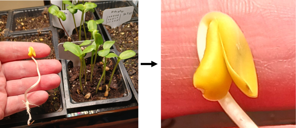
    One of the easiest ways to “make” etioplasts at home is to grow seedlings in the dark. In Figure 1, you can see a sunflower seedling grown in the dark, side-by-side with seedlings grown in the light. These seedlings are all the same age, but there are some notable differences! For starters, the dark-grown seedling is a lot smaller. Furthermore, if we zoom in and look at the cotyledons (embryonic structures resembling leaves), we can see that they are yellow in the dark-grown seedling and green in the light-grown seedlings. This yellow coloration indicates the presence of etioplasts!

    Figure 1. Left: Dark-grown sunflower seedling (in my hand) compared to light-grown sunflower seedlings of the same age. Right: A close-up of the cotyledons of the dark-grown seedling. They contain lots of etioplasts!

    Ok, great! Can we see some etioplasts under the microscope?

    … *sigh*. I tried. I really did. But I encountered some difficulties in doing so. The main problem is that the cotyledon cells in the dark-grown seedling are very small and densely packed together. Light attenuates quickly in such densely-packed tissue, making it difficult to visualize on the microscope (I just see dark-colored blobs). Fine. So next, I tried squishing the tissue in the hopes of splitting a few individual cells off to look at. But I found that the cells were very… crumbly. When I squished the tissue, the cells just ruptured and released their contents in a giant maelstrom of crap (See Figure 2 below). Some of the little globules you can see are probably etioplasts, but since they are not darkly pigmented, it is impossible to differentiate them from other cell contents.

    Figure 2. The big mess made by squishing tissue from dark-grown cotyledons.

    One possible explanation for the brittle-ness of the dark-grown cotyledon cells could be that their cell walls are thinner than in light-grown seedlings. I couldn’t find information about cotyledons specifically, but I did find this source which shows that cell wall thickness increases in the hypocotyls (stems)of sunflower seedlings after they are exposed to light [2]. This makes intuitive sense, since plants with access to light can photosynthesize and obtain more carbon to build cell walls with.

    Wherefore art thou, chloroplast?

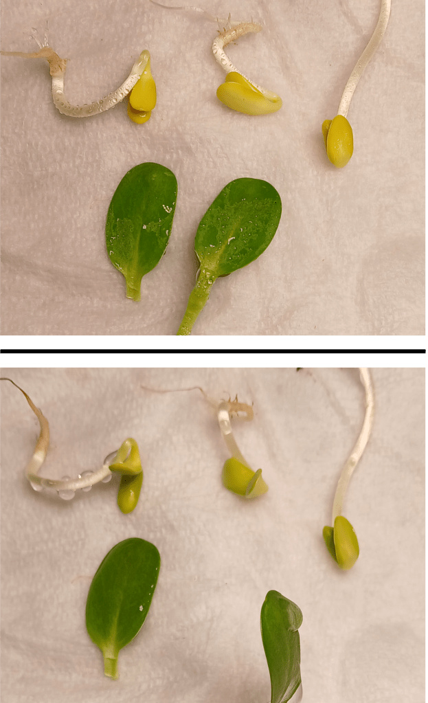
    OK, so we can’t easily look at etioplasts directly. However, we can observe the etioplast-to-chloroplast transition by looking at whole seedlings! As I mentioned before, etioplasts become chloroplasts when they are exposed to light. We can see this change by looking at the color change of dark-grown cotyledons, from yellow to green! For an example, see Figure 3 below. This color change occurs because light stimulates production of chlorophyll, the green pigment required for photosynthesis.

    Figure 3. Dark-grown seedlings vs. two dissected cotyledons from a light-grown seedling. Top: The dark-grown seedlings were just transferred to light. Bottom: The dark-grown seedlings were exposed to light for 8 hours.

    How do etioplasts sense light, and how does this lead to chlorophyll production? I’m glad you asked! Etioplasts contain an important enzyme called protochlorophyllide oxidoreductase (POR for short) [3]. This enzyme catalyzes a key step in chlorophyll biosynthesis. I was surprised to learn that the enzyme itself is light-activated – no other upstream signaling mechanisms are needed [3]! Very cool.

    What would happen if a plant were deficient in the POR enzyme? As you might expect, Arabidopsis mutants with reduced levels of POR have reduced chlorophyll content and various other chloroplast defects [1]. Arabidopsis actually has 3 POR genes. Single mutants don’t have an obvious phenotype, but double mutants do [1]. The presence of multiple genes which can perform the same tasks is known as genetic redundancy. In the case of POR, the porB porC double mutant is seedling lethal – that is, plants cannot grow beyond the seedling stage before dying [1].

    Mixed results:

    We’re all scientists here. And scientists measure things! So…. Is there a way to quantify the greening process?

    Well, sure. You could extract and quantify the concentration of chlorophyll in seedlings – but this requires a bunch of specialized equipment that I don’t have. However, I did come across this interesting paper which proposes a way to estimate chlorophyll content from RGB photographs, such as those you can take with a smartphone camera [4]! In particular, they found that the ratio of green to red pixel channel values was a reasonably good proxy for chlorophyll content [4]. This makes intuitive sense if you consider that in RGB, green color is created by having a high green channel value, while yellow is made by mixing red and green channels together. A low G:R ratio gives yellow and indicates low chlorophyll content, while a high G:R ratio gives green and indicates a high chlorophyll content!

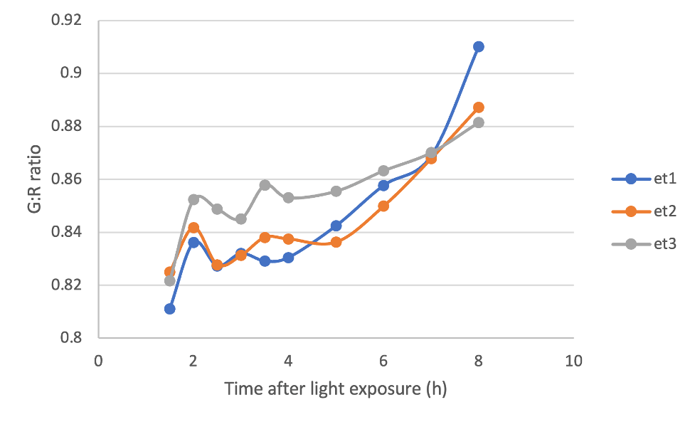
    With this in mind, I photographed cotyledons from 3 different dark-grown seedlings after they were exposed to light, and quantified the G:R ratio of the pixels making up these cotyledons using an image-processing software called FIJI. Below, you can see that the G:R ratio for my three seedlings increases with time as they were exposed to light, indicating accumulation of chlorophyll. It works!

    Figure 4. G:R ratio of rectangular selections of cotyledons from 3 dark-grown seedlings over time after they were exposed to light.

    It… er… might have been a bit too early to celebrate. I also took the G:R ratio of a couple of light-grown cotyledons. We would expect these values to be relatively constant over time, since the chlorophyll content of mature cotyledons shouldn’t change meaningfully over only a few hours. However, if we plot the G:R ratio of light-grown cotyledons on the same graph as our dark-grown cotyledons, we get something that looks like Figure 5 below.

    Figure 5. G:R ratio of rectangular selections of cotyledons from 3 dark-grown seedlings and 2 light-grown cotyledons over time.

    What to heck?! The G:R ratios of the light-grown cotyledons are super variable! But why? Troubleshooting is an important part of the scientific process, so let’s think of some possibilities.

    One issue that I found was that the surfaces of the light-grown cotyledons were somewhat reflective under my lights, especially when water droplets were present on their surface. Why were there water droplets? Since the cotyledons no longer had access to water, I was misting them with water constantly to keep them moist and alive. Unfortunately, this probably also had an effect on my photographs.

    Another interesting thing that I noticed was that the blue channel values for pixels from the dark-grown cotyledons were virtually 0 throughout the entire experiment. However, the blue channel values for pixels from the light-grown cotyledons were higher and quite variable. In instances where the blue channel values were high, the green value channels tended to be lower, indicating a hidden relationship between the blue and green channels. It is possible that this “hidden relationship” made my results more variable. I don’t know exactly why this occurred. One interesting possibility is that this is due to the presence of other pigments (such as anthocyanins) in the light-grown seedlings, but I cannot say for certain.

    I’m definitely going to try this again, with an improved protocol. Stay tuned.

    Gene of the week:

    Well alrighty, it’s that time again! This week’s genes of the week will be the POR genes. As previously mentioned, Arabidopsis has 3 POR genes, which act redundantly with each other to catalyze chlorophyll production in etioplasts undergoing the transition to chloroplasts [1].

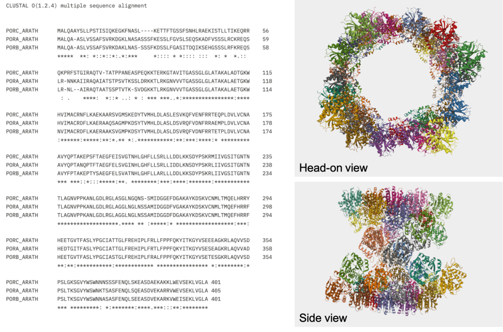
    All of the POR proteins in Arabidopsis are roughly 400 amino acids long and have a mass of around 43 kDa (according to their respective UniProt entries). If we align their sequences using Clustal Omega (https://www.ebi.ac.uk/jdispatcher/msa/clustalo), we can see that their sequences are extremely similar (See Figure 6) – indicating that the 3 genes probably emerged from a recent duplication event.

    But it gets crazier! I found this interesting paper which uses a technique called cryo-electron microscopy to look at the structure of POR [5]. They found that POR proteins come together to form enormous tubular structures! Apparently, these tubes help to organize internal membranes within the plastid [5]. And they look oh-so-cool! See Figure 6 below for an example of the structure.

    Figure 6. Left: Sequence alignment of the 3 POR proteins from Arabidopsis. Right: Structure of PORB. The sequences and the structure were obtained from the following UniProt entries:

    https://www.uniprot.org/uniprotkb/Q42536/entry#sequences

    https://www.uniprot.org/uniprotkb/P21218/entry#sequences

    https://www.uniprot.org/uniprotkb/O48741/entry#sequences

    Works Cited:

    [1] Frick, G., Su, Q., Apel, K., & Armstrong, G.A. (2003). An Arabidopsis porB porC double mutant lacking light-dependent NADPH:protochlorophyllide oxidoreductases B and C is highly chlorophyll-deficient and developmentally arrested. The Plant Journal 35(20, 141-153.

    [2] Kutschera, U. (1990). Cell-wall synthesis and elongation in the hypocotyls of Helianthus annuus L. Planta 181, 316-323.

    [3] Menon, B.R.K., Davidson, P.A., Hunter, C.N., Scrutton, N.S., & Heyes, D.J. (2009). Mutagenesis Alters the Catalytic Mechanism of the Light-driven Protochlorophyllide Oxidoreductase. Journal of Biological Chemistry 285(3), 2113-2119.

    [4] Nasoha, N.Z., Ibrahim, N.U.A., Harith, H.H., Jamaludin, D., & Abd Aziz, S. (2025). Linear regression and machine learning modelling for chlorophyll content estimation using leaf red, green, and blue images. Food Research 9(1), 94-100.

    [5] Nguyen, H.C., Melo, A.A., Kruk, J., Frost, A., & Gabruk, M. (2021). Photocatalytic LPOR forms helical lattices that shape membranes for chlorophyll synthesis. Nature Plants 7, 437-444.

  • How do plants sense gravity?

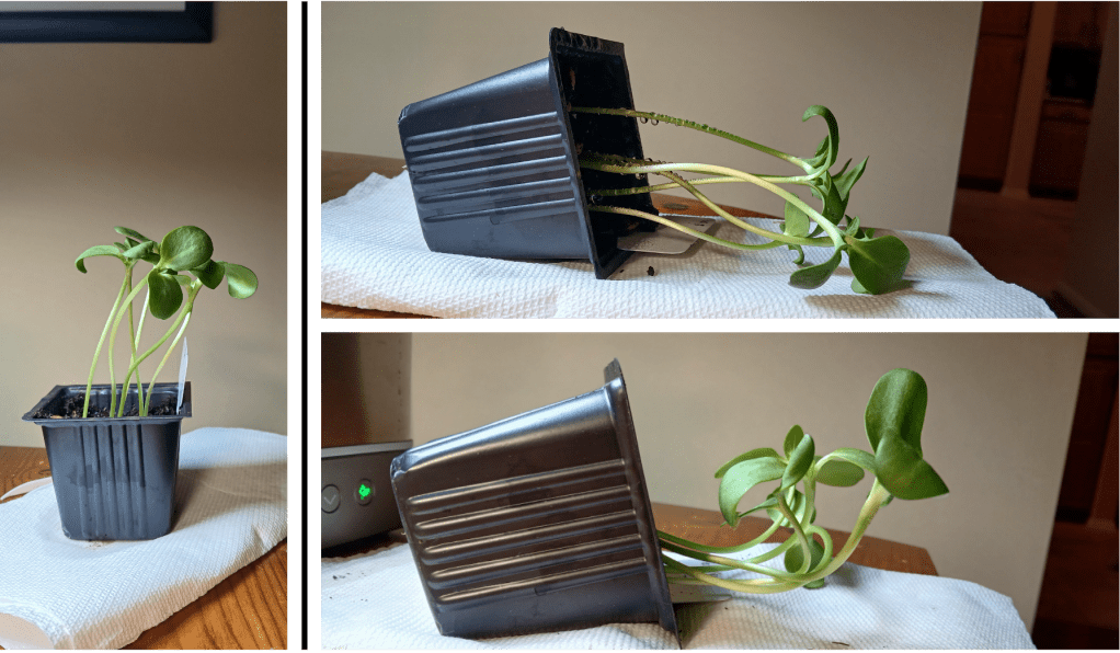
    I’ve been growing some sunflower seedlings indoors under artificial lights. To demonstrate that plants can sense gravity, I turned one pot on its side and left it in a dark cupboard overnight. As you can see in Figure 1, the seedlings reorient their growth so that they once again face upward. But how do they know which way the gravity is going??

    The answer is specialized plastids called amyloplasts, which contain large quantities of starch (“amlyo” is Greek for starch). We covered the concept of plastids in last week’s post. In short, plastids are organelles (subcellular structures) which have their own DNA and which live inside of plant cells. They can take on a number of different identities and may become chloroplasts, chromoplasts, amyloplasts, etc. Plants sense gravity using specialized amyloplasts called statoliths (Greek for “standing stone”, because they look like little stones! You’ll see why in a moment).

    Figure 1. Left: sunflower seedlings used to test gravity-sensing. Right top: The same pot turned on its side. Right bottom: the same pot after sitting on its side in the dark overnight. One seedling was removed for microscopy (you’ll see why in a moment!)

    Let’s see some statoliths!

    Okey dokey! Let’s see some statoliths! But how?

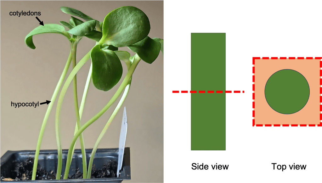
    We’ll need to look at cells in the hypocotyl of our sunflower seedlings. The hypocotyl is the stem of the seedling (See Figure 2 left), which is derived from an embryonic structure that forms early in seedling development. Note that roots also have statoliths and can sense gravity independently of the stem… but I will discuss those in a later blog post.

    Anyway, statoliths in the model plant Arabidopsis thaliana are known to be found in the endodermal cell layer in stems [7]. Before going any further, I wanted to check whether the same was true for sunflower hypocotyls. To do this, I took a horizontal section through the hypocotyl (See Figure 2 right) and had a look under the microscope.

    Figure 2. Left: Sunflower seedlings, showing the location of the hypocotyl and cotyledons (leaf-like structures).

    The endodermis is a layer of cells that surrounds the pith and vascular tissue. In an unstained section of hypocotyl, it is extremely difficult to make out (See Figure 3 top left). Fortunately, we have some Lugol’s iodine at our disposal! You might remember from a science class at some point that iodine stains starch. Using Lugol’s iodine to stain our hypocotyl sections allows us to see the location of our statoliths – and by extension, the endodermis. You can see a hypocotyl section stained with Lugol’s iodine in the top right of Figure 3. A thin line of black spots belies the position of the endodermis. If you look even more closely, you can see individual statoliths within the endodermal cells (Figure 3 bottom).

    Figure 3. Top left: Unstained section of sunflower hypocotyl at 100X magnification. Top right: Section of sunflower hypocotyl stained with Lugol’s iodine at 100X magnification. The position of the endodermis is indicated. Bottom: Endodermis and surrounding tissues at 400X magnification. Individual statoliths are visible.

    Statoliths facilitate gravity sensing because they are denser than other cell contents and sink to the bottom of cells [7]. More on that in a minute. But this means that if we want to see statoliths in action, we need to take a vertical section of hypocotyl and view cells from the side. (See Figure 4 top for the location of the cuts I made). When we look at these vertical sections without staining, it is difficult to identify the endodermis cells (Figure 4 bottom left). However, after staining with Lugol’s iodine, the location of the statoliths/endodermis becomes clear (Figure 4 bottom right)!

    Figure 4. Top: Locations of cuts to make to take vertical sections of hypocotyl tissue. Bottom left: Unstained hypocotyl section, with visible tissues labelled. Bottom right: Hypocotyl section stained with Lugol’s iodine. The endodermis, which contains statoliths, is visible.

    Statolith sedimentation:

    As previously mentioned, statoliths are believed to facilitate gravity sensing because they sink to the bottom of the cells. Sinking of the statoliths sets off a signaling cascade which ultimately results in a redistribution of the growth hormone auxin within the stem/hypocotyl, which leads to asymmetric cell elongation, which in turn allows plants to change the direction of their growth [5][6]. Exactly how the statolith sensing mechanism works is still being intensively studied. The review article by Kawamoto & Morita [4] provides an excellent summary of several lines of research, but we still don’t fully understand how we get from statoliths sinking –> auxin redistribution.

    Er… right. Given that we don’t understand precisely how statolith-mediated gravity sensing works, how do we even know that these statoliths are important at all?? Fortunately, there is good evidence that points in this direction. For example, Arabidopsis mutants which lack the endodermal cell layer entirely are agravitropic (do not respond to gravity) [3]. Furthermore, Arabidopsis mutants which lack starch-filled amyloplasts, such as pgm (phosphoglucomutase) mutants, have a weaker gravitropic response (though it is not entirely gone) [2].

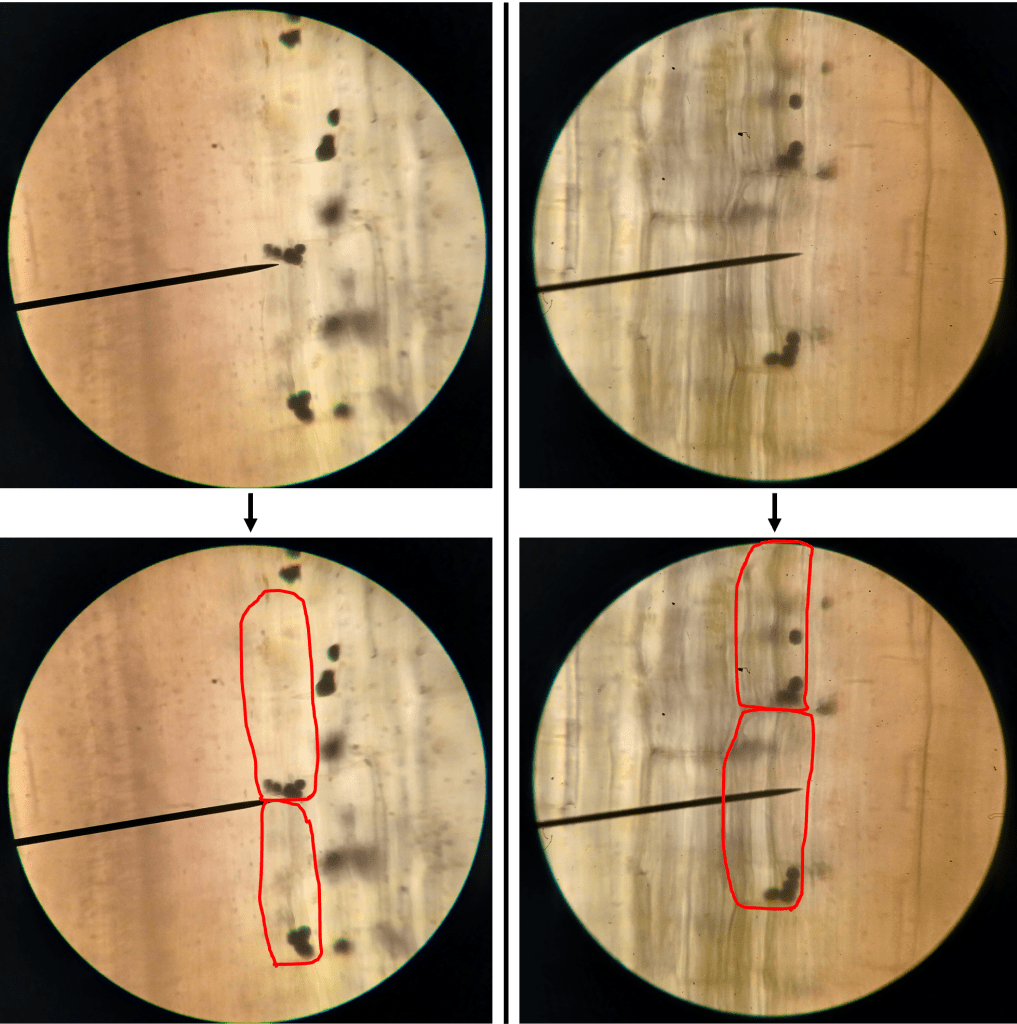
    All very cool. But can we see statolith sedimentation for ourselves? Sure we can! First, I looked at statoliths in a vertical section of hypocotyl in a seedling that was growing vertically upwards. You can see that in Figure 5 that the statoliths accumulate on the bottom of the cells, as expected. Since the cell boundaries are difficult to see, I’ve highlighted them in red.

    Figure 5. Statoliths in sunflower hypocotyls in a seedling which was growing vertically. Top: Unaltered images. Bottom: Images with cell boundaries highlighted for clarity.

    Very good. Now, what happens if we tilt a seedling onto its side, wait a few hours, and then look at the statoliths? See for yourself in Figure 6! Here, statoliths accumulate on the lower side of the cells, as expected.

    Figure 6. Statoliths in sunflower hypocotyls in a seedling which was tilted horizontally for several hours. Top: Unaltered images. Bottom: Images with cell boundaries highlighted for clarity.

    Gene of the week:

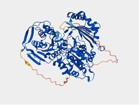
    And just like that, it’s time for gene of the week! This week’s gene will be PGM (phosphoglucomutase) in Arabidopsis. PGM codes for an enzyme which is essential for the biosynthesis of starch in amyloplasts. As previously noted, pgm mutants have a reduced gravitropic response [2]. As expected, the UniProt database entry (https://www.uniprot.org/uniprotkb/Q9SCY0/entry#subcellular_location) notes that the PGM protein localizes to plastids. It also tells us that the PGM gene in Arabidopsis is located at the AT5G51820 locus on chromosome 5, and codes for a protein which is 623 amino acids long, with a mass of ~68 kDa. There is a nice alphafold structure available:

    Figure 7. Alphafold structure of Arabidopsis PGM, taken from its UniProt entry.

    I did a quick search of the OMA database (https://omabrowser.org/oma/home/) to see if there are any known orthologs of Arabidopsis PGM in sunflowers. For genes involved in such essential biochemical pathways, I would expect to see lots of orthologs with a high degree of sequence similarity across many species, so this should be a breeze. Indeed, OMA tells us that a predicted sunflower protein called “HELAN14524” exists, which has a similar sequence to Arabidopsis PGM. Furthermore, it is possible to predict that the sunflower PGM protein also localizes to plastids, because it contains a Transit Peptide sequence – a protein sequence motif that acts kind of like a “mailing address” to send proteins to plastids [1].

    Works cited:

    [1] Bruce, B.D. (2000). Chloroplast transit peptides: structure, function, and evolution. Trends in Cell Biology 10(10), 440-447.

    [2] Caspar, T., & Pickard, B.G. (1989). Gravitropism in a starchless mutant of Arabidopsis. Planta 177, 185-197.

    [3] Fukaki, H., Wysocka-Diller, J., Kato, T., Fujisawa, H., Benfey, P.N., & Tasaka, M. (2002). Genetic evidence that the endodermis is essential for shoot gravitropism in Arabidopsis thaliana. The Plant Journal 14(4), 425-430.

    [4] Kawamoto, N., & Morita, M.T. (2022). Gravity sensing and responses in the coordination of the shoot gravitropic setpoint angle. New Phytologist 236, 1637-1654.

    [5] Rakusová, H., Abbas, M., Han, H., Song, S., Robert, H.S., & Friml, J. (2016). Termination of Shoot Gravitropic Responses by Auxin Feedback on PIN3 Polarity. Current Biology 26(22), 3026-3032.

    [6] Wang, X., Yu, R., Wang, J., Lin, Z., Han, X., Deng, Z., Fan, L., He, H., Deng, Z.W., & Chen, H. (2020). The Asymmetric Expression of SAUR genes Mediated by ARF7/19 Promotes the Gravitropism and Phototropism of Plant Hypocotyls. Cell Reports 31(2), 107529.

    [7] Wyatt, S.E., Rashotte, A.M., Shipp, M.J., Robertson, D., & Muday, G.K. (2002). Mutations in the Gravity Persistence Signal Loci in Arabidopsis Disrupt the Perception and/or Signal Transduction of Gravitropic Stimuli. Plant Physiology 130(3), 1426-1435.